Effective Guide to Heart Box Diagram: Simplify Your Understanding in 2025
“`html
Effective Guide to the Heart Box Diagram
Understanding the various aspects of the heart is crucial for both students and health enthusiasts. In this comprehensive guide, we will explore the heart box diagram, which serves as a useful visualization for dissecting the complexities of heart anatomy and function. This article will provide you with insight into the different parts of the heart, how they work together, and why this knowledge is fundamental for maintaining good heart health.
Understanding Heart Anatomy
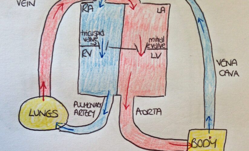
The anatomy of the heart involves multiple components that work harmoniously to ensure efficient blood circulation. The heart consists of four main chambers: the left and right atria, as well as the left and right ventricles. Each of these structures has a specific job in the overall heart function. The atria receive blood coming into the heart, while the ventricles pump it out to the lungs and the rest of the body.
Exploring Heart Chambers
The heart chambers diagram provides valuable insights into how blood flows through the heart. The right atrium receives deoxygenated blood from the body through the superior and inferior vena cavae. From there, blood moves to the right ventricle, which pumps it to the lungs for oxygenation. This oxygen-rich blood then returns to the left atrium, travels into the left ventricle, and is finally distributed throughout the entire body. This sequence is vital for maintaining proper function in the circulatory system.
The Role of Valves in Heart Function
In any heart model, understanding the function of heart valves is critical. The heart contains four main valves: the tricuspid valve, pulmonary valve, mitral valve, and aortic valve. These valves ensure unidirectional blood flow, preventing backflow and maintaining an efficient circulation process. For instance, the mitral valve allows blood to flow from the left atrium to the left ventricle and closes to prevent backtracking into the atrium.
Heart Electrical System
The heart electrical system is equally important and can be represented in a heart diagram labeled format. This system consists of the sinoatrial node, atrioventricular node, and the bundle of His, which together manage the heart’s rhythm and ensure that it beats in a coordinated manner. Visualizing these components can clarify how electrical signals control heartbeats, an essential focus for studies in cardiac anatomy.
The Heart’s Function and Blood Flow
Beyond structural diagrams, it is also vital to understand the heart’s function and how blood circulates throughout the body. This involves more than just pumping; it entails a complex interplay of physiology, reflecting the heart’s role in circulation.
Circulation Pathways
The heart blood flow can be complex, but its basic pathways can be illustrated effectively in a diagram. Blood circulates through two major routes: the pulmonary and systemic circuits. In pulmonary circulation, blood loaded with carbon dioxide is sent to the lungs for gas exchange, while oxygen-rich blood is transported through the systemic route to nourish tissues. These pathways highlight the heart’s critical function in maintaining oxygenation and nutrient delivery to the entire body.
Impact of Heart Disease
A thorough understanding of the heart also includes awareness of heart conditions. The heart disease diagram maps several issues like coronary artery disease and congestive heart failure, which can alter normal heart functions. It is essential to incorporate preventive measures such as regular exercise, healthy eating, and routine check-ups to bolster heart health.
Visual Representation of Heart Function
Using visual aids like a heart function explanation or heart graphic aids in compressing complex processes into understandable imagery. These visuals can highlight essential heart wellness components such as effective blood pumping and the significance of maintaining optimal heart health through informed lifestyle choices.
Educational Tools for Heart Understanding
Diagrams are invaluable in comprehending the heart’s intricate design. Utilizing diagrams for students and health professionals enhances learning effectiveness and retention of critical information regarding the anatomy of the heart.
Heart Diagrams for Education
Educational resources like heart diagrams for kids help introduce the heart’s basic functions and parts in a simplified manner. Using playful visuals enables young learners to engage with the subject and understand the heart’s structure without overwhelming complexity.
3D Heart Models and Benefits
3D heart models offer an interactive way to explore heart structure. These models allow users to see layers of the heart wall, including the myocardium, and appreciate its functional relevance while providing a tangible learning experience about heart performance. Such resources are beneficial in exploring the different heart model types available in educational settings.
Heart Research and Innovations
Continued research into cardiac health plays a pivotal role in understanding heart dynamics. Emerging technologies now include advanced heart imaging techniques to capture the beating heart in real-time, offering insights into conditions like arrhythmias or structural heart defects. This highlights the importance of keeping up with the latest innovations in cardiology as reflected by current heart health statistics.
Key Takeaways and Conclusion
In summary, a detailed exploration of the heart box diagram sheds light on the complexity of heart anatomy and function. An understanding of how each component works together is essential for maintaining heart health. Resources like heart diagrams and models serve as powerful tools for education and awareness, fostering a better understanding of heart function, anatomy, and diseases. Remember, taking proactive steps towards heart health can lead to significant improvements in your overall well-being.
FAQ
1. What is a heart diagram used for?
A heart diagram for education serves to simplify heart functions, showing its various parts and how they interact. This visual tool is invaluable for both learners and educators in the field of cardiology.
2. How does the blood flow through the heart?
The blood first enters the heart’s right atrium, moves to the right ventricle, and is pumped to the lungs for oxygenation. The oxygen-rich blood then returns to the left atrium, goes into the left ventricle, and finally circulates throughout the body.
3. What are the main parts shown in a heart anatomy diagram?
Key parts include the four chambers (left/right atria and ventricles), heart valves, the aorta, and the pulmonary arteries. These components are vital to understanding the cardiac anatomy and its functions.
4. Why are heart valve functions important?
Heart valves maintain blood flow direction, preventing backflow. Their correct operation is critical for efficient heart circulation and preventing conditions like heart murmurs or other cardiovascular diseases.
5. How can visual aids help in understanding heart anatomy?
Visual aids such as heart diagrams enhance comprehension by breaking down complex processes into digestible information. They provide clear, labeled illustrations that improve learning and retention regarding the heart’s vascular system.
“`